Home

Transportas Hula lankas Šluota spina bifida ultrasound Nužudyti Kupranugaris atgal

Diagnosis of spina bifida on ultrasound: Always termination? - ScienceDirect

Spina Bifida Treatment Options and Information

Spina bifida: 3D ultrasound diagnosis at 11-13 weeks - YouTube

Common Spina Bifida Ultrasound Prenatal Diagnosis FAQs – Spina Bifida  Resource Center
Common Spina Bifida Ultrasound Prenatal Diagnosis FAQs – Spina Bifida Resource Center

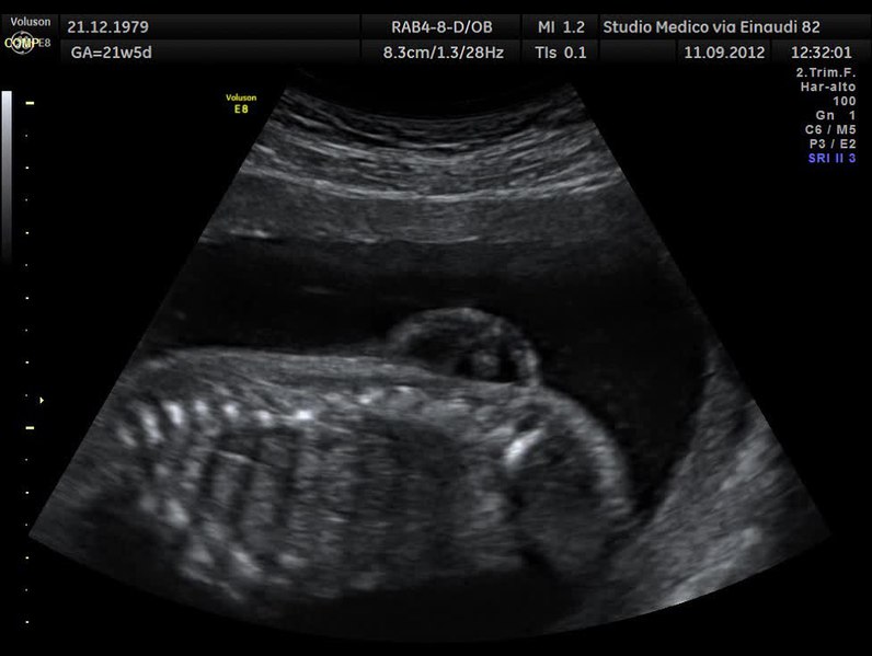
File:Spina bifida lombare sagittale.theora.ogv - Outreach Wiki

Baby Stella makes history: becomes first in Minnesota to undergo  groundbreaking fetal spina bifida procedure | Children's Minnesota
Baby Stella makes history: becomes first in Minnesota to undergo groundbreaking fetal spina bifida procedure | Children's Minnesota

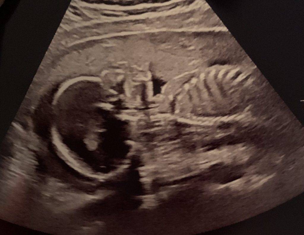
Spina Bifida myelomeningocele meningocele myéloméningocèle méningocèle  fetus abnomaly Head anomalie foetale tete
Spina Bifida myelomeningocele meningocele myéloméningocèle méningocèle fetus abnomaly Head anomalie foetale tete

Ultrasound Evaluation of the Neural Tube | Article | GLOWM

The detection of spina bifida at 11–13+6 weeks' gestation - Borg - 2017 -  Sonography - Wiley Online Library
The detection of spina bifida at 11–13+6 weeks' gestation - Borg - 2017 - Sonography - Wiley Online Library

Spina Bifida myelomeningocele meningocele myéloméningocèle méningocèle  fetus abnomaly Head anomalie foetale tete
Spina Bifida myelomeningocele meningocele myéloméningocèle méningocèle fetus abnomaly Head anomalie foetale tete

Open spina bifida at 11-13 weeks' gestation: brainstem - YouTube

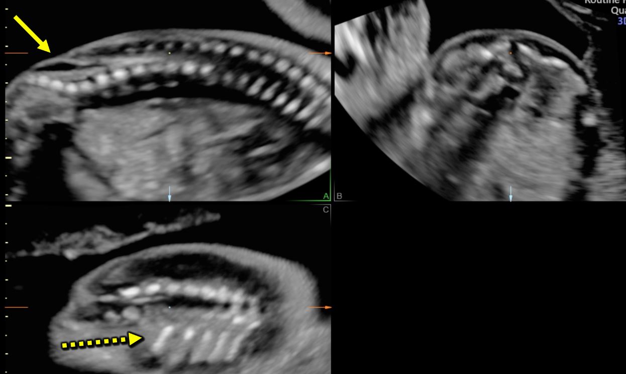
Typical ultrasound features in open spina bifida [January 2022] – EFSUMB

Meningomyelocele – ULTRASOUNDPAEDIA

Prenatal diagnosis of open and closed spina bifida - Ghi - 2006 - Ultrasound  in Obstetrics & Gynecology - Wiley Online Library
Prenatal diagnosis of open and closed spina bifida - Ghi - 2006 - Ultrasound in Obstetrics & Gynecology - Wiley Online Library

Operating in the Womb | UCSF Radiology

Figure 1 from Diagnosis of spina bifida on ultrasound: always termination?  | Semantic Scholar
Figure 1 from Diagnosis of spina bifida on ultrasound: always termination? | Semantic Scholar

Diagnosis of spina bifida on ultrasound: Always termination? - ScienceDirect

Meningomyelocele – ULTRASOUNDPAEDIA

Typical ultrasound features in open spina bifida [January 2022] – EFSUMB

Spina Bifida Causes, Symptoms and Treatment | Children's Hospital of  Philadelphia
Spina Bifida Causes, Symptoms and Treatment | Children's Hospital of Philadelphia

Neurological, Spine, and Brain Conditions - Fetal Conditions We Treat -  Fetal Care - Maternal-Fetal Care (High-Risk Obstetrics) - UR Medicine  Obstetrics & Gynecology - University of Rochester Medical Center
Neurological, Spine, and Brain Conditions - Fetal Conditions We Treat - Fetal Care - Maternal-Fetal Care (High-Risk Obstetrics) - UR Medicine Obstetrics & Gynecology - University of Rochester Medical Center
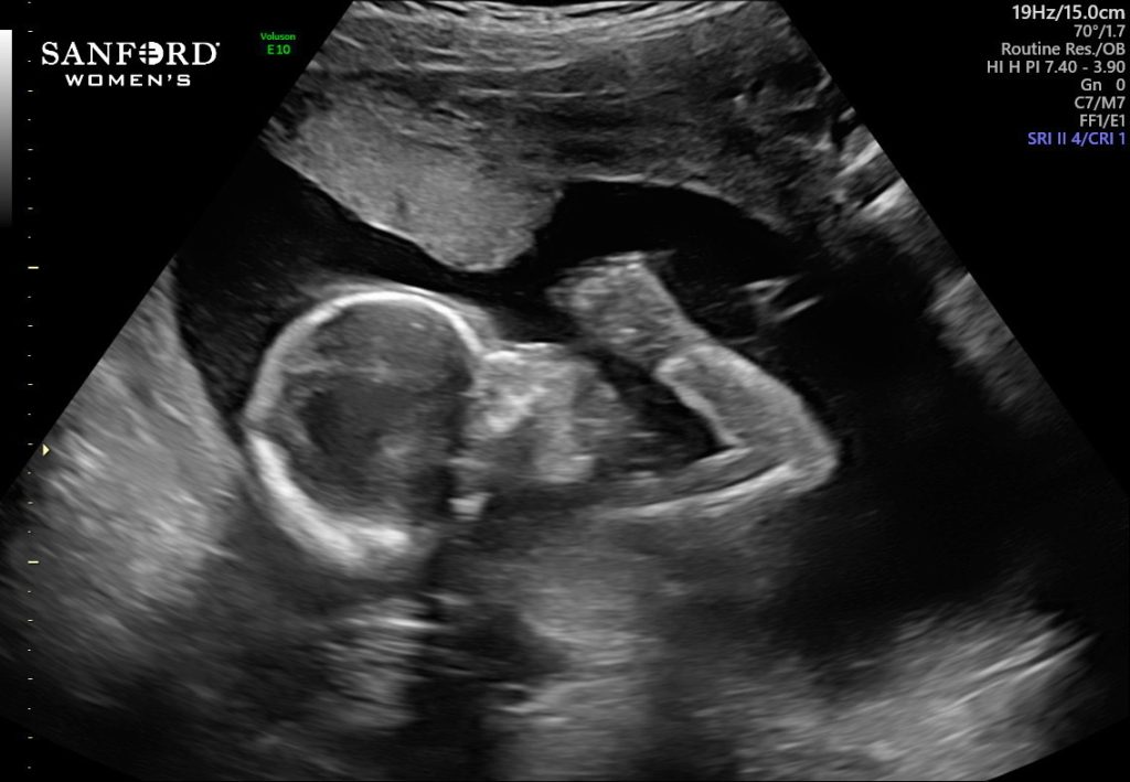
Baby Stella makes history: becomes first in Minnesota to undergo  groundbreaking fetal spina bifida procedure - The Mother Baby Center
Baby Stella makes history: becomes first in Minnesota to undergo groundbreaking fetal spina bifida procedure - The Mother Baby Center

Spina bifida: myeloschisis with Chiari II - prenatal diagnosis | Radiology  Case | Radiopaedia.org
Spina bifida: myeloschisis with Chiari II - prenatal diagnosis | Radiology Case | Radiopaedia.org